Silent vision thief glaucoma develops normal intraocular pressure...Who's at risk?
Mar 09, 2025
According to statistics from the Health Insurance Review and Assessment Service, the number of patients treated for glaucoma in 2023 is 1.19 million. This is a 20% increase from 970,000 in 2019, showing a continuous increase over the past five years.
Glaucoma is an ophthalmic disease that causes visual field problems due to optic nerve damage and is one of the leading causes of blindness around the world. Glaucoma causes glaucoma optic papillary changes and corresponding visual field deficits due to various factors such as intraocular pressure and blood flow. Except for cases such as acute obstructive angle glaucoma, there are often no or insignificant symptoms in the early stages, making it difficult to recognize visual field damage.
Jang Yoon-kyung, head of the ophthalmology department at Bundang Jesaeng Hospital, said, `In the early stages of glaucoma, the peripheral vision narrows very slightly, but at the end of glaucoma, the vision narrows as if viewed through a tunnel, eventually leading to blindness. In addition, in the case of acute obstructive angle glaucoma, severe pain or rapid vision loss may occur because the intraocular pressure rises rapidly. If you feel headache and eye pain as the front of your eyes turns blurry, or if you feel symptoms of dangling around you when you look at the light, you should see an ophthalmologist as soon as possible.."
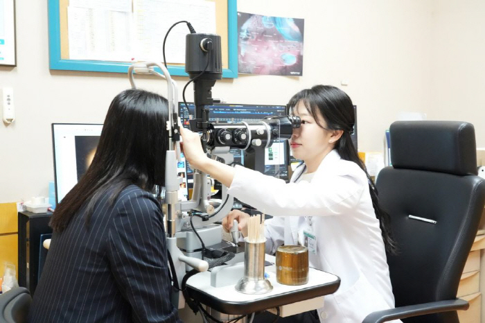
Glaucoma test methods include intraocular pressure measurement, corneal thickness measurement, optic nerve examination, and visual field examination. High intraocular pressure is one of the most important risk factors for glaucoma, and corneal thickness is an important factor to increase the accuracy of intraocular pressure measurement, and the thicker the cornea, the higher the intraocular pressure can be measured than the actual intraocular pressure. The optic nerve examination is conducted by directly identifying the optic nipple through fundus examination, or using fundus photography, optic fiber layer photography, and light interference tomography (OCT), and determines whether and how damaged the optic nerve is. The visual field examination determines visual field damage due to glaucoma by measuring visual field range and sensitivity. In addition, angiography, municipal development gastroscopy, and genetic testing are used to diagnose and determine whether glaucoma progresses.
Since the optic nerve once damaged by glaucoma does not recover, the main goal of glaucoma treatment is to prevent the progression of optic nerve damage and prevent blindness. Currently, the main method in the treatment of glaucoma is intraocular pressure control. An intraocular pressure lowering agent and eye drops are used as the primary treatment to lower intraocular pressure. If sufficient intraocular pressure drop is difficult with the drug, laser treatment or surgical treatment may be required. Recently, treatment has been underway in the direction of dealing with systemic abnormalities such as blood circulation disorders as well as intraocular pressure, and improving the quality of life of glaucoma patients.
Director Jang Yoon-kyung said, `Although the increase in intraocular pressure is the main cause of glaucoma, there are many cases of normal intraocular pressure glaucoma in Korea that occur even though the intraocular pressure is within the normal range. This is because the degree of intraocular pressure that the optic nerve can withstand varies from individual to individual and is normal intraocular pressure, but if the optic nerve is weak, it can be easily damaged. Until optic nerve damage progresses above a certain level, most people find glaucoma during medical checkups because they do not feel vision loss or visual impairment, and if they are aware of glaucoma symptoms, they are often quite advanced. Therefore, it is most important to detect and treat glaucoma early.'
Since only a portion of all ophthalmic diseases can be detected with basic medical checkups, if you are over 40 years old or have other diseases such as myopia, family history of glaucoma, intraocular pressure, diabetes, high blood pressure, and cardiovascular disease, you should have an ophthalmologic examination every year.
Glaucoma is an ophthalmic disease that causes visual field problems due to optic nerve damage and is one of the leading causes of blindness around the world. Glaucoma causes glaucoma optic papillary changes and corresponding visual field deficits due to various factors such as intraocular pressure and blood flow. Except for cases such as acute obstructive angle glaucoma, there are often no or insignificant symptoms in the early stages, making it difficult to recognize visual field damage.
Jang Yoon-kyung, head of the ophthalmology department at Bundang Jesaeng Hospital, said, `In the early stages of glaucoma, the peripheral vision narrows very slightly, but at the end of glaucoma, the vision narrows as if viewed through a tunnel, eventually leading to blindness. In addition, in the case of acute obstructive angle glaucoma, severe pain or rapid vision loss may occur because the intraocular pressure rises rapidly. If you feel headache and eye pain as the front of your eyes turns blurry, or if you feel symptoms of dangling around you when you look at the light, you should see an ophthalmologist as soon as possible.."
Glaucoma test methods include intraocular pressure measurement, corneal thickness measurement, optic nerve examination, and visual field examination. High intraocular pressure is one of the most important risk factors for glaucoma, and corneal thickness is an important factor to increase the accuracy of intraocular pressure measurement, and the thicker the cornea, the higher the intraocular pressure can be measured than the actual intraocular pressure. The optic nerve examination is conducted by directly identifying the optic nipple through fundus examination, or using fundus photography, optic fiber layer photography, and light interference tomography (OCT), and determines whether and how damaged the optic nerve is. The visual field examination determines visual field damage due to glaucoma by measuring visual field range and sensitivity. In addition, angiography, municipal development gastroscopy, and genetic testing are used to diagnose and determine whether glaucoma progresses.
Since the optic nerve once damaged by glaucoma does not recover, the main goal of glaucoma treatment is to prevent the progression of optic nerve damage and prevent blindness. Currently, the main method in the treatment of glaucoma is intraocular pressure control. An intraocular pressure lowering agent and eye drops are used as the primary treatment to lower intraocular pressure. If sufficient intraocular pressure drop is difficult with the drug, laser treatment or surgical treatment may be required. Recently, treatment has been underway in the direction of dealing with systemic abnormalities such as blood circulation disorders as well as intraocular pressure, and improving the quality of life of glaucoma patients.
Director Jang Yoon-kyung said, `Although the increase in intraocular pressure is the main cause of glaucoma, there are many cases of normal intraocular pressure glaucoma in Korea that occur even though the intraocular pressure is within the normal range. This is because the degree of intraocular pressure that the optic nerve can withstand varies from individual to individual and is normal intraocular pressure, but if the optic nerve is weak, it can be easily damaged. Until optic nerve damage progresses above a certain level, most people find glaucoma during medical checkups because they do not feel vision loss or visual impairment, and if they are aware of glaucoma symptoms, they are often quite advanced. Therefore, it is most important to detect and treat glaucoma early.'
Since only a portion of all ophthalmic diseases can be detected with basic medical checkups, if you are over 40 years old or have other diseases such as myopia, family history of glaucoma, intraocular pressure, diabetes, high blood pressure, and cardiovascular disease, you should have an ophthalmologic examination every year.
|
This article was translated by Naver AI translator.