With 3D and virtual dyeing technology, cancer tissue can be observed without incision
May 26, 2025
|
|
The Korea Advanced Institute of Science and Technology (KAIST) announced that a team of physics professors Park Yong-geun developed an innovative technology that vividly shows the three-dimensional structure of cancer tissues without dyeing separately from Shin Soo-jin of Yonsei University Gangnam Severance Hospital, Hwang Tae-hyun of Mayo Clinic in the U.S., and Tomocube's artificial intelligence research team. Park Joo-yeon, a master's and doctoral integration student at KAIST, participated as the first author and was published online on the 22nd in Nature Communications, a world-renowned academic journal.
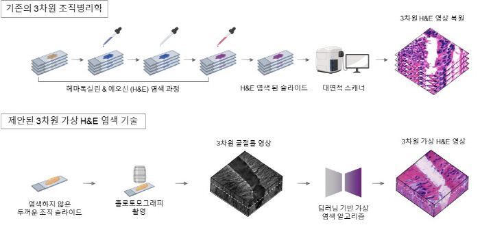
In existing pathology, which has been used for more than 200 years, the method of observing cancer tissue under a microscope only shows a specific cross-section of cancer tissue in three dimensions, so there was a limit to grasping the three-dimensional connection structure or spatial arrangement between cells.
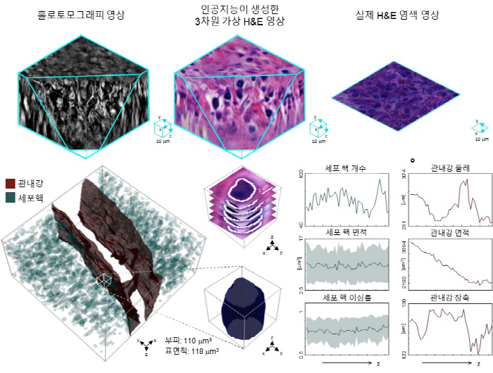
In response, the research team used advanced optical technology called 'Holotomography (HT)' to measure the tissue's three-dimensional refractive index information and combine artificial intelligence-based deep learning algorithms to generate virtual H&E images like real errors. Hematoxylin & Eosin (H&E) is the most widely used staining method when observing pathological tissue, and the nucleus of the cell is blue with hematoxylin and the cytoplasm is pink with Eosin.
The research team explained that it quantitatively proved that the images produced by the technology were very similar to actual stained tissue images, and proved versatility and reliability as a next-generation pathology analysis tool by showing consistent performance in various organs and tissues.
Professor Park Yong-geun said "This study is a very meaningful achievement of expanding the analysis unit of pathology from two dimensions to three dimensions" and "In the future, it will be widely used for various biomedical research and clinical diagnosis, such as analyzing the spatial distribution of cancer tumor boundaries or surrounding marginal cells within the microtumor environment."
This article was translated by Naver AI translator.