Development of the world's first simultaneous nerve and tumor identification imaging technique...New Standard for Precision Surgical Surgery
Aug 06, 2025
|
During tumor surgery, it is difficult to determine the exact location of the nerve due to changes in the anatomical structure, and there is a high risk of nerve damage. In particular, if the laryngeal, penile, and sympathetic nerves present around the thyroid, esophagus, and prostate are damaged, they can cause serious aftereffects such as postoperative voice changes, swallowing disorders, sexual dysfunction, and sensory degradation. Previously, electrophysiological tests, ultrasound, and optical coherence tomography (OCT) were used, but there were limitations in terms of real-time performance, resolution, and visibility.
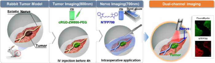
To overcome these limitations, the research team developed and utilized two fluorescent contrast agents that responded to each wavelength in the near-infrared band, and established the world's first technology to visualize nerves and tumors simultaneously during surgery.
The research team newly developed a nerve-specific fluorescent contrast agent 'NTFP700' and implemented a dual-channel near-infrared (NIR) imaging technique that visualizes nerves as blue fluorescence at 700 nm wavelength and tumors as green fluorescence at 800 nm wavelength using the existing tumor target contrast agent 'cRGD-ZW800-PEG'. The technique was applied to a rabbit sciatic nerve perioperative tumor model and succeeded in simultaneously identifying tumors and nerves during surgery. For tumor labeling, cRGD-ZW800-PEG was intravenously injected 4 hours before surgery and observed with 800 nm channel, and nerve labeling was applied with 700 nm channel during surgery by adsorbing NTFP700 onto gauze.
As a result, the tumor and nerve were clearly separated at the surgical site, so that the resection boundary could be set and the nerve preservation could be determined in real time. The fluorescence signal was also consistent with the biopsy results after resection, demonstrating the high targetability and reproducibility of the imaging technique. In particular, by clearly visualizing tumors and nerves in different colors through fluorescent cameras, the possibility of implementing a surgical environment that can precisely resect tumors while minimizing nerve damage was confirmed.
In particular, the research team newly introduced a dyed gauze method that absorbs the fluorescent contrast agent NTFP700 into the sterile gauze and covers the nerve area directly. Existing direct application methods had limitations in that the contrast medium flowed in the direction of gravity or the dyeing was uneven in anatomically complex areas, but the dyeing gauze method uniformly covered the nerves to enable constant and stable fluorescent dyeing. In particular, nerve contrast was clearly secured in inclined structures and curved areas, and it was confirmed that the surgeon could easily apply it during surgery, satisfying both practicality and accuracy.
Professor Choi Hak-soo of Harvard Medical School said, "This is an example of a new solution that can improve the accuracy of surgery and maximize patient safety. In particular, dual-channel imaging techniques that can separate and visualize nerves and tumors in real time have the potential to fundamentally change the scene of surgery beyond simple technological development."."
Professor Kim Hyun-gu of Cardiovascular Thoracic Surgery at Korea University Guro Hospital said, "Nerve damage that occurs during cancer surgery is not just a side effect, but a serious threat to the patient's vocal, sensory, and motor functions, as well as the quality of life as a whole." "This study proved the world's first technology to separate and visualize nerves and tumors in real time during surgery at the preclinical stage, which marked an important turning point in improving both precision of surgery and patient safety."," he said.
Meanwhile, the study was recently published in ACS (Applied Materials & Interfaces, IF: 8.2), a prestigious international academic journal in materials science and biotechnology, under the title 'Tumor-Embedded Nerve Survey Using Dual-Channel Interoperative Imaging Programs' (Tumor-Embedded Nerve Survey).
|

This article was translated by Naver AI translator.