Severance Hospital's first successful diagnosis and treatment of cerebrospinal fluid venous fistula...All four patients with brain haemorrhages who couldn't find the cause recovered
Oct 16, 2025
|
Cerebral cerebrospinal fluid venous fistula is one of the rare forms of spontaneous intracranial hypovolemia, in which cerebrospinal fluid that protects the brain and maintains brain pressure (intacranial pressure) leaks into the veins around the spinal cord through 'fistula (fistula)'. It occurs without a clear cause and is difficult to diagnose, but as a result, it lowers the pressure on the brain, causing various symptoms.
Standing headaches, which worsen headaches when standing up, cognitive impairment that reduces judgment or problem-solving ability, and walking disorders have a big impact on daily life.
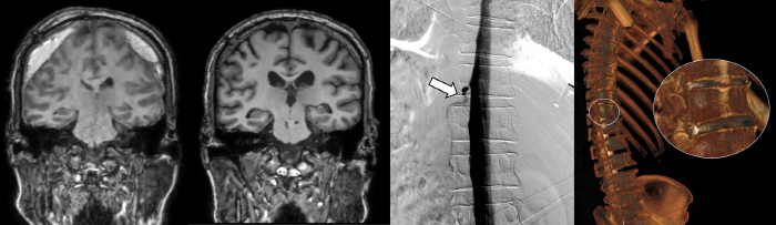
Unlike common spontaneous intracranial low pressure, the subtype, cerebrospinal fluid venous fistula, is a disease that has difficulty from identifying the cause to treatment because it can show normal findings in MRI.
In fact, all four patients with cerebrospinal fluid venous fistula treated this time were suspected of having 'spontaneous intracranial hypotension', which causes headaches due to low brain pressure before coming to Severance Hospital, but both spinal MRI and simple spinal angiography showed normal findings.
This is why symptoms did not improve even if self-blood removal was performed to prevent the leak of cerebrospinal fluid with one's blood. In addition, the bridge vein connecting the inside of the dura mater surrounding the brain and the vein was cut off due to the low pressure brain, which also caused subdural hemorrhage, making it impossible to walk on its own.
Severance Hospital recently introduced DSM for the first time in Korea to overcome the limitations of such existing diagnostic methods. Digital Subtraction Myelography (DSM) can inject contrast agents into the spinal cord and check the flow of cerebrospinal fluid in real time with a monitor screen. Through this, it is possible to accurately find holes through which cerebrospinal fluid leaks.
Along with DSM, positional CT spinal cord angiography, which is being conducted by Severance Hospital, is all the latest techniques to diagnose cerebrospinal fluid venous fistula.
As neurologists (Ju Min-kyung, Professor Ha Woo-seok) and neurosurgeons (Professor Ha Yoon) work closely together, diagnosis and treatment are one-stop.
Patients who visited Severance Hospital this time were accurately diagnosed and treated with DSM tests and positional CT spinal cord angiography, restored cerebral pressure, and disappeared subdural hemorrhage. As a result, both cognitive decline and walking disorders seen by patients improved.
Severance Hospital said the preemptive introduction of multidisciplinary treatment and 3D real-time testing methods, in which several medical specialists consider treatment strategies for each patient, is the basis for successful diagnosis and treatment of cerebrospinal fluid venous fistula in Korea for the first time.
Professor Ha Woo-seok said, `The cerebrospinal fluid spinal fistula, which was one of the causes of spontaneous intracranial pressure reduction and sub-dural hemorrhage without special trauma, causes serious pain such as headaches and cognitive decline, but it was not easy to determine the cause with conventional diagnostic methods.' `DSM and positioning CT spinal cord angiography introduced by Severance Hospital can accurately identify and treat the location of spinal fluid leakage.'
|

This article was translated by Naver AI translator.