3D patent technology enables actual closed state...Development of Lung Nodulation Position Visualization Thoracic Simulation
May 20, 2025
|
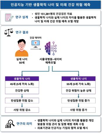
Professor Hwang's team recently completed domestic patent registration by developing a thoracoscopic simulation device based on a three-dimensional inorganic lung model.
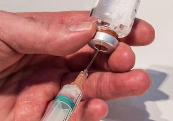
Chest CT is taken at the maximum level of inhalation, but actual surgery is performed in a lung-depleted state with no air. For this reason, it is difficult to find the exact location of the lung nodule during surgery because the CT image and the actual appearance of the lungs are different, so the location of the lung nodule is found through invasive procedures such as dyeing and radioactive material injection. Professor Hwang believed that these methods could pose additional risks to patients, such as continuous radiation exposure and incision of abnormal tissue, and developed the technology to improve stability and surgical accuracy.
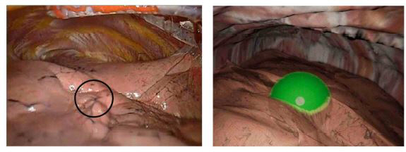
This patent technology is a system that creates an inorganic lung 3D model similar to the actual surgical environment based on CT images of patients and visualizes the location and resection range of lung nodules and outputs them as images. It is a principle that mathematically simulates the intake 3D model obtained from CT images by reflecting gravity and posture changes, and analyzes the location of the ribs around it to accurately reproduce the lung shape during actual surgery. You can check the preoperative simulation image and compare it with the actual thoracoscopic image during surgery.
Professor Hwang explained "Using this technique, we can accurately predict the actual location and extent of resection of lung nodules in advance." In addition, "As the internal structure of the lungs can be checked in advance before surgery, it is possible to improve the safety and success rate of surgery with an accurate resection plan as well as shortening the operation time."
|

This article was translated by Naver AI translator.