Abdominal aortic aneurysm with more than 60% mortality in case of rupture, and a clue to treatment was found
Sep 03, 2025
|
An international joint research team consisting of Professor Oh Se-jin of Cardiovascular Thoracic Surgery at Boramae Hospital in Seoul, Professor Kim Eun-na of the Department of Pathology at Seoul National University Hospital, Professor Jang Young-hwan of Oregon Health Science University, and Dr. Lizhe Zhuang of Cambridge University in the U.K. sought the answer by analyzing the immune cell microenvironment in abdominal aortic aneurysm tissue.
What the research team paid attention to is an inflammation-related substance called C-reactive protein (CRP). In particular, we investigated how CRP in the form of monomer (mCRP) adhering to the vascular wall changes immune cell composition at the single cell level, among others.
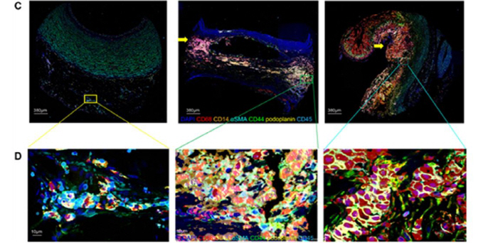
To this end, the research team divided patients into groups with high CRP deposition (High-CRP) and low-CRP (Low-CRP) based on the patients' serum CRP levels and the degree of staining in tissues. And patient tissues were stained and analyzed with 31 antibodies using a high-resolution imaging technique called 'CODEX'. This allowed us to check hundreds of thousands of cells at once, determine which immune cells and matrix cells are in them, and how they are located.
As a result, in the High-CRP group with high CRP deposits, inflammation-causing M1 macrophages and highly proliferative macrophages increased, and smooth muscle cells that make up blood vessels were noticeably reduced. In addition, the spatial relationship between immune cells also changed, such as regulatory T cells clustered close to NK cells, B cells, and endothelial cells.
On the other hand, in the Low-CRP group, fibrosis was more prominent than inflammation, and M2 macrophages with anti-inflammatory action were relatively common. Overall, it was confirmed that the more CRP deposits, the more inflammatory cells and the fewer the matrix cells that make up the tissue.
Thanks to CODEX technology, the research team was able to analyze not only the type of each cell but also how close the cells were to each other. In particular, high-resolution images showed a dense structure in which regulatory T cells, NK cells, and macrophages were clustered in the center of the atherosclerotic lesion.
This study is the first to precisely show how the composition and spatial arrangement of inflammatory cells vary depending on the degree of CRP deposition in patients with abdominal aortic aneurysm. The research team expects that if CRP can act as a key immune regulator that affects disease progression, it will lead to the development of new treatment strategies targeting it in the future.
Professor Oh Se-jin of Boramae Hospital said, "This is the first time we have specifically confirmed how the composition of immune cells in the abdominal aortic aneurysm varies depending on the degree of deposition of C-reactive proteins." It was revealed.
The study was carried out with the support of the Korea Research Foundation's Personal Basic Research Project Excellent Progress Research Project, and the results of the study were recently published in the international journal 『Translational Research』.
|

This article was translated by Naver AI translator.